Salivary Gland Tumors and Most Common Types
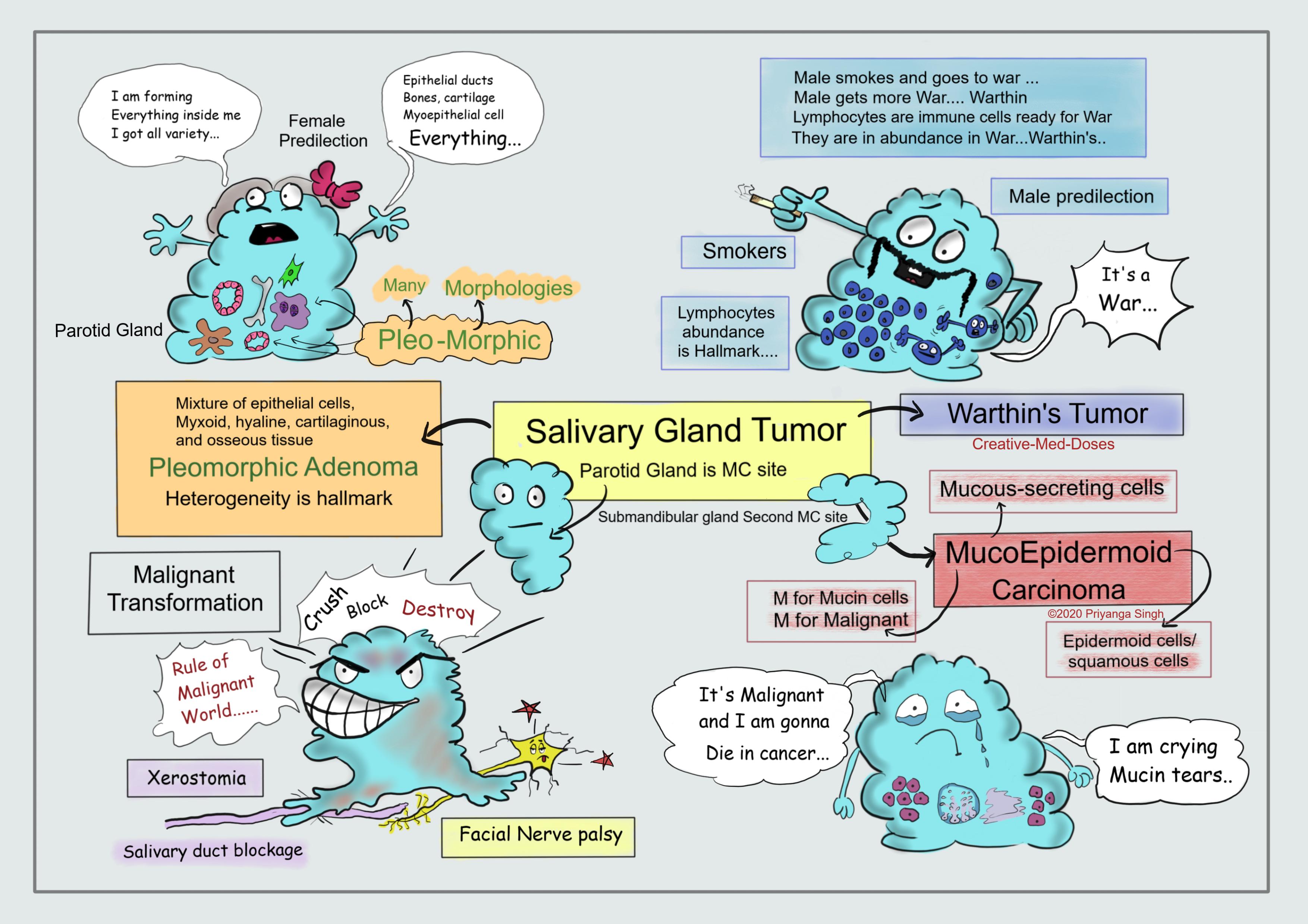
Salivary gland tumors are rare tumors that begin in the salivary glands. Salivary gland tumors can begin in any of the salivary gland, but parotid gland is most common site.
Following are the most common forms-
Pleomorphic Adenoma
- Pleomorphic adenomas are benign tumors consisting of mixture of epithelial and mesenchymal cells.
- Epithelial elements are dispersed in the matrix, which contain variable mixtures of myxoid, hyaline, cartilaginous, and osseous tissue.
- Because of this histologic diversity it is also called mixed tumor.
- Pleomorphic adenomas are most common benign tumors of the parotid gland.
- They present as slow-growing, painless, mobile discrete masses. It can recur if incompletely excised.
- Pleomorphic adenomas typically manifest as rounded, well demarcated masses rarely exceeding 6cm in the greatest dimension. They are encapsulated but sometimes expansile growth produces protrusions into the surrounding tissues such as facial nerve and salivary ducts.
- On cutting, gross examination shows gray-white areas mixed with myxoid and blue translucent cartilage-like areas. The most striking histologic feature is heterogeneity. Epithelial elements resembling ductal or myoepithelial cells are typically are dispersed within a mesenchyme-like background of loose myxoid tissue containing islands of chondroid and, occasionally foci of bone.
- Carcinoma arising in a pleomorphic adenoma is referred to variously as a carcinoma ex pleomorphic adenoma or malignant mixed tumor. The incidence of malignant transformation increases with time.
Mucoepidermoid Carcinoma
- Mucoepidermoid carcinomas are composed of variable mixtures of squamous cells, mucus-secreting cells, and intermediate cells.
- Most common Malignant tumor of salivary gland.
- They occur mainly in (60%–70%) the parotids.
- Mucoepidermoid carcinomas lack well-defined capsules and often are infiltrative.
- The cut surface is pale gray to white and frequently demonstrates small, mucinous cysts.
- Biopsy from these tumors contain cords, sheets, or cysts lined by squamous, mucous, or intermediate cells. The cysts are lined by mucous cells and filled with mucin and it can be appreciated by mucin stains.
Warthin's tumor
- Benign
- second most common benign tumor of parotid (first is pleomorphic adenoma)
- more common in males
- contains abundant lymphocytes and germinal center
- associated with smoking
...
...
Clinical presentation of Most Salivary neoplasm is similar-
- Lump and swelling in affected gland, mostly painless.
- Xerostomia
If malignant ---
- Facial nerve palsy
- Fixation of lump on overlying skin and ulceration/ induration of involved mucosa.
Diagnosis-
- CT or MRI to determine location and extent of tumor
- Fine Needle Aspiration Cytology
- Biopsy
Treatment
Surgical resection of the tumor (if tumor is malignant lymph nodes are also removed).
Case scenario
A 65-year-old woman presents with history of a painless lump over jaw region that has been there for 4 years. Recently, she has noticed increment in size, she also complains of facial drooping with slurring of speech. Physical exam reveals firm, non-tender mass. The neck lymph nodes are not palpable. The biopsy of lump reveals epithelial cells dispersed in the matrix, which contain mixtures of myxoid, hyaline, cartilaginous, and osseous tissue. Which is most likely diagnosis?
A. Mucoepidermoid Carcinoma
B. Osteomyelitis mandible
C. Pleomorphic Adenoma
D. Facial nerve tumor
Interesting video https://www.youtube.com/watch?time_continue=389&v=AQvjJdZJuSU&feature=emb_logo
Revision for today https://creativemeddoses.com/topics-list/irritable-bowel-syndrome-stress-and-diarrhea/