Renal artery stenosis and Secondary hypertension
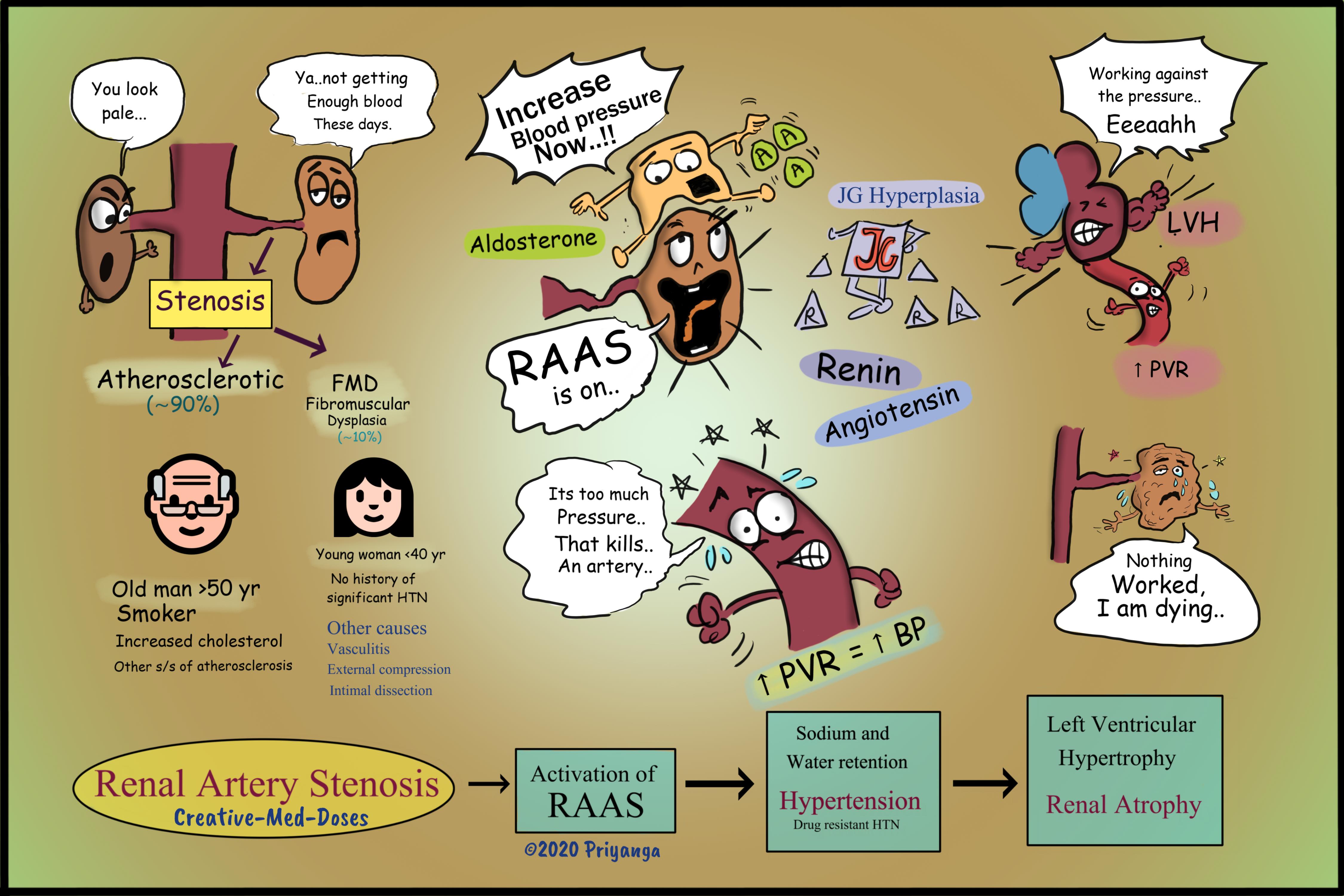
Renal artery stenosis is the narrowing of one or both renal arteries, which leads to hypertension and renal atrophy.
Etiology
Atherosclerosis
The majority of cases (nearly 90%) are associated with lumen narrowing due to atherosclerotic plaque, which occurs more in men > 50 years of age. Atherosclerotic RAS (ARAS) typically involves the proximal third of the renal artery including the perirenal aorta and ostium. In these patients, atherosclerosis is generally systemic and not limited to the renal artery, and elderly patients present with multiple cardiovascular risk factors (hyperlipidemia, smoking, history of stroke, etc).
Fibromuscular dysplasia (FMD)
There is a thickening of the renal arterial wall causing luminal narrowing in (nearly 10% of cases). It mostly affects women < 40 years of age. FMD has classically been described as having a “beads-on-a-string” appearance due to contrast filling of consecutive aneurysms along the renal artery. In fibromuscular dysplasia, the muscle and fibrous tissues in the arterial wall thickening, causing the arteries to narrow. This may reduce blood flow to the kidney, leading to kidney damage and Renovascular hypertension.
Other causes include vasculitis, external compression, and radiation therapy.
Pathophysiology
Renal artery stenosis/luminal narrowing → reduced renal perfusion and slow pressure in renal vasculature → activation of RAAS (renin-angiotensin-aldosterone system) → Increased renin and aldosterone → Increased sodium and water reabsorption → Sodium and water retention → volume expansion → Increased peripheral vascular resistance → Renovascular hypertension (secondary hypertension).
Long-standing renal hypoperfusion → continuous stimulation of the juxtaglomerular apparatus to secrete renin → juxtaglomerular apparatus hyperplasia
Ischemic nephropathy
Severe reduction in renal blood flow → ischemia of renal tissue → renal insufficiency, cortical thinning and progressive renal atrophy →Chronic kidney disease and renal failure
Left ventricular hypertrophy and pulmonary edema
Increased Peripheral vascular resistance → increased afterload → left ventricular hypertrophy →increased left ventricular filling pressure → reduced ejection fraction → pulmonary edema
...

...
Clinical presentation
- Hypertension which doesn’t respond to usual antihypertensive medicine
- Deterioration in renal function after taking an ACE inhibitor or angiotensin receptor blocking (ARB) agent
- heart failure or pulmonary edema
Diagnosis
- Duplex Ultrasonography
- CT or MR angiography
- Invasive Catheter Angiography (gold standard)
Treatment
Antihypertensive medicine
ACE-inhibitors or ARBs
- Given with precautions, and regular serum creatinine level checkups
- Contraindicated in B/LRenal stenosis and patient with a single kidney
Calcium channel blockers or beta-blockers in patients not responding to ACE inhibitors, or where it is contraindicated.
Surgical intervention
Revascularization
Reserved for cases which don’t respond to medical treatment
- Balloon dilation of the stenosis
- Stent placement
Revision for today https://creativemeddoses.com/topics-list/gitelman-syndrome-ncct-defect/
Follow us on Facebook https://www.facebook.com/creativemeddoses
Buy fun review books here (these are kindle eBook’s you can download kindle on any digital device and login with Amazon accounts to read them). Have fun and please leave review.