IgA nephropathy: Berger disease
IgA nephropathy /Berger disease is the glomerulonephritis which is characterized by episodic hematuria and deposition of IgA in the mesangium (the hallmark of the disease). IgA nephropathy is the most common form of glomerulonephritis in adults and it affects males in the second to third decades of life (male preponderance).
It can occur sporadically or secondary to the liver or intestinal illness. In liver disease clearance of IgA is impaired which increases IgA levels in the blood. The intestinal illness increases IgA levels by increased mucosal production of IgA.
Pathogenesis
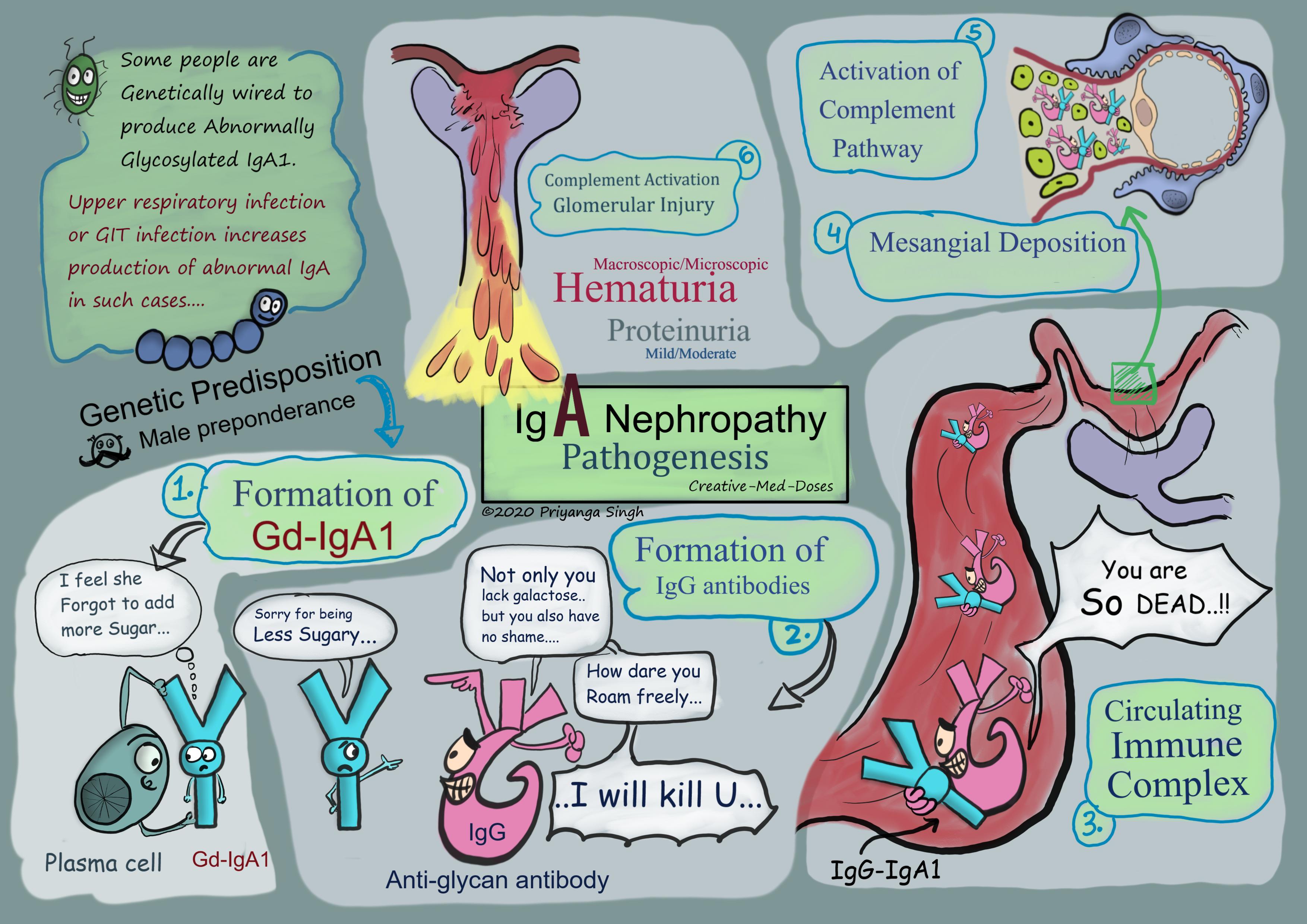
The exact mechanism is still unclear. Following steps are thought to be related to IgA nephropathy-
- Formation of abnormally Glycosylated IgA1
An abnormally glycosylated IgA1 (i.e., galactose-deficient IgA1 [Gd-IgA1]) immunoglobulin has a central role in the pathogenesis. Why some people produce defective IgA is genetically decided. In these genetically susceptible individuals, respiratory or gastrointestinal exposure to microbial or other antigens (e.g., viruses, bacteria, food proteins such as gluten in celiac disease) may lead to increased IgA synthesis, some of which is abnormally glycosylated.
- Formation of antibody against abnormally glycosylated IgA1 (anti-glycan antibodies)
The accumulated abnormal IgA stimulate autoimmune response, and IgG autoantibodies are formed. These IgG autoantibodies form large immune complexes by binding with circulating IgA.
- Formation of circulating immune complexes
The abnormally glycosylated IgA1 and IgG antibodies form immune complexes and are circulating in blood.
- Deposition of immune complexes at mesangium
These complexes deposit in the glomerular mesangium; this unusual location may be related to physicochemical features of the IgA and may be facilitated by an IgA1 receptor (CD71) on mesangial cells.
- Glomerular injury
The presence of C3 in the mesangium and immune complex deposited on mesangium activates the alternative complement pathway and initiate glomerular injury. The hematuria and proteinuria occur due to the injured glomeruli filtration membrane.
...

...
HSP and IgA nephropathy
Clinical and laboratory features show close similarities between Henoch-Schönlein purpura and IgA nephropathy. IgA nephropathy and HSP both are IgA mediated diseases triggered by a mucosal infection. Henoch-Schonlein purpura is distinguished clinically from IgA nephropathy by having the following differences-
- More prominent systemic symptoms in HSP in contrast to IgA nephropathy which affects kidney predominantly
- Younger age of presentation (<10 years old), IgA nephropathy typically affects adults.
- Abdominal complaints like diarrhea and crampy pain are characteristic in HSP
To Read more on HSP, click here 👉 Henoch-Schonlein purpura
Clinical presentation
The two most common presentations of IgA nephropathy are recurrent episodes of macroscopic hematuria following an upper respiratory or GIT infection often accompanied by proteinuria, or persistent asymptomatic microscopic hematuria which remains asymptomatic in nearly 30% of cases.
The course of the disease is highly variable and can manifest in the following patterns -
- Asymptomatic cases that have proteinuria and microscopic hematuria but remain asymptomatic. They are diagnosed when they come for a routine checkup.
- Recurring episodic illness presenting with
- Gross or microscopic hematuria
- Flank pain
- Low-grade fever
- Nephritic syndrome (including hypertension and proteinuria)
- Rarely presents with RPGN and/or nephrotic syndrome
Diagnosis
Clinical presentation and laboratory results help in making diagnosis, gross hematuria following upper respiratory tract infection or intestinal infection is an important clue for diagnosis.
Urinalysis and microscopy
- Hematuria (gross or microscopic)
- Proteinuria
- RBC cast
Laboratory tests
- Serum IgA level is elevated
- Complement levels are normal
Renal biopsy findings
Light microscopy shows mesangial proliferation and mesangial widening.
Immunofluorescent microscopy reveals mesangial IgA deposits.
Electron microscopy: mesangial immune complex deposits.
Treatment
Patients with isolated hematuria normally recover spontaneously, monitoring kidney function and initiate treatment if disease progresses (patient gets proteinuria or hypertension).
Patients with proteinuria or hypertension and hematuria -ACE inhibitors/angiotensin II receptor blockers
For severe and rapidly progressive disease: (nephrotic range proteinuria and hypertension)
Glucocorticoids and cyclophosphamide/azathioprine is given. Plasmapheresis is reserved for refractory cases.
Prognosis
Most important factor in prognosis is magnitude, duration and qualitative aspects of proteinuria.
Renal biopsy findings which are related with poorer prognosis-
- diffuse mesangial proliferation
- segmental sclerosis
- endocapillary proliferation
- tubulointerstitial fibrosis.
A 30-year-old male presents to his physician with hematuria 6 days after the onset of a productive cough, sore throat and fever. The renal biopsy immunofluorescence shows granular IgA deposits in the glomerular mesangium. Which of the following is most likely disease?
- Poststreptococcal glomerulonephritis
- Berger Disease
- Alport syndrome
- Minimal Change Disease
Answer is B
Revision for today Acute Pyelonephritis
Buy fun review books here (these are kindle eBook’s you can download kindle on any digital device and login with Amazon accounts to read them). Have fun and please leave review. https://creativemeddoses.com/books/