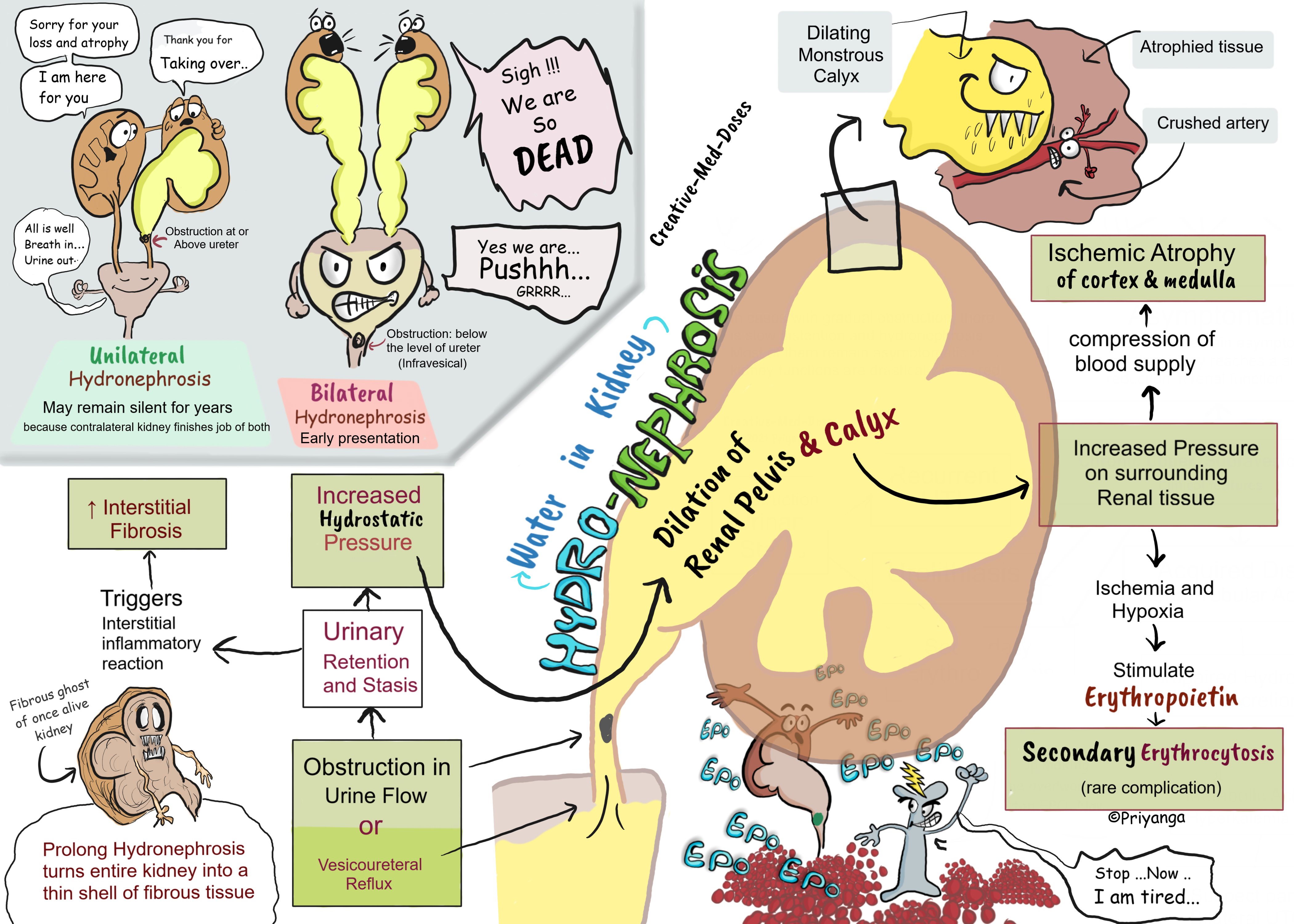
Hydronephrosis: Dilated Pelvis and calyx
Hydronephrosis means dilation of renal calyx and pelvis. It occurs due to an obstruction in urine flow or vesicoureteral reflux leading towards urinary retention and stasis. The urinary tract obstruction may be sudden or insidious.
Following are the most common causes
- Congenital causes
- atresia of the urethra,
- valve formations in either the ureter or urethra
- an aberrant renal artery compressing the ureter
- renal ptosis with torsion
- kinking of the ureter
- Acquired causes
- Calculi or sloughed necrotic papillae
- Proliferative lesions, such as benign prostatic hyperplasia, carcinoma of the prostate, bladder tumors,
- Contiguous malignant disease retroperitoneal lymphoma, and carcinoma of the cervix or uterus
- Inflammatory lesions, such as prostatitis, ureteritis, urethritis, and retroperitoneal fibrosis
- Neurogenic, such as paralysis of the bladder following spinal cord damage
- Normal pregnancy (enlarged uterus compressing the urinary tract and obstructing the urinary outflow)
Bilateral hydronephrosis occurs only when the obstruction is below the level of the ureters (Infravesical Obstruction).
If the blockage is at the ureters or above, there will be unilateral hydronephrosis.
Pathogenesis
Obstruction of urinary flow → urinary retention in renal pelvis and calyces → increased hydrostatic pressure in collecting system → dilation of the renal pelvis and calyces → distention of the renal pelvis and calyces → increased pressure on the surrounding renal parenchyma → interrupted blood supply of surrounding renal tissue → causing ischemia and loss of renal parenchyma→atrophy of Cortex and the Medulla
The most severe effects are seen in the papillae because they are vulnerable to even the slightest of ischemia.
The initial functional disturbances are mainly tubular, causing impaired urine concentrating ability and polyuria.
The obstruction and urinary retention trigger an interstitial inflammatory reaction → increase recruitment of inflammatory cells and fibroblasts → interstitial fibrosis.
Bilateral hydronephrosis or unilateral hydronephrosis with a solitary kidney leads to renal failure and uremia.
Depending on the level of the obstruction one, or both ureters also may be dilated (hydroureter).
Morphology
The cut section of the hydronephrotic kidney shows dilated renal pelvis and calyces and thinned cortex of the kidney.
Microscopy
the early lesions show tubular dilation, followed by atrophy and fibrous replacement of the tubular epithelium.
In severe cases, the glomeruli also become atrophic and lesser in number.
Prolonged hydronephrosis may convert the entire kidney into a thin shell of fibrous tissue.
...
...
Clinical Features
Depend on whether the obstruction is acute or chronic and unilateral or bilateral
Most cases are asymptomatic an enlarged kidney is discovered on routine physical examination (incidental finding).
Bilateral complete obstruction produces anuria. The dominant symptoms are those of bladder distention in cases with bladder outlet obstruction.
The incomplete bilateral obstruction causes polyuria instead of oliguria. Impaired tubular urine concentrating ability and tubular atrophy cause polyuria in the early stages of incomplete bilateral obstruction.
Unilateral hydronephrosis may remain completely silent for long periods unless the other kidney also is dysfunctional.
Serum creatinine elevates only when there is a bilateral obstruction or if the patient has a solitary kidney.
Physical examination findings
- palpable kidney in severe cases
- costovertebral angle tenderness
- distended bladder in lower urinary tract obstruction (e.g., benign prostatic hyperplasia)
Diagnosis
Renal ultrasonography- initial investigation of choice to detect the extent of dilation and site of obstruction
CT scan of the abdomen- used when USG doesn’t give much information or non-confirmatory
Treatment
Treat the underlying cause.
If the patient presents with signs and symptoms of Bladder neck Obstruction, perform bladder catheterization to relieve the pressure.
Prognosis depends upon the underlying cause, the extent of distention, and the amount of renal atrophy.
Revision for today Restrictive Cardiomyopathy: Stiff Ventricles - Creative Med Doses
Buy fun review books here (these are kindle eBook’s you can download kindle on any digital device and login with Amazon accounts to read them). Have fun and please leave review.
https://creativemeddoses.com/books/