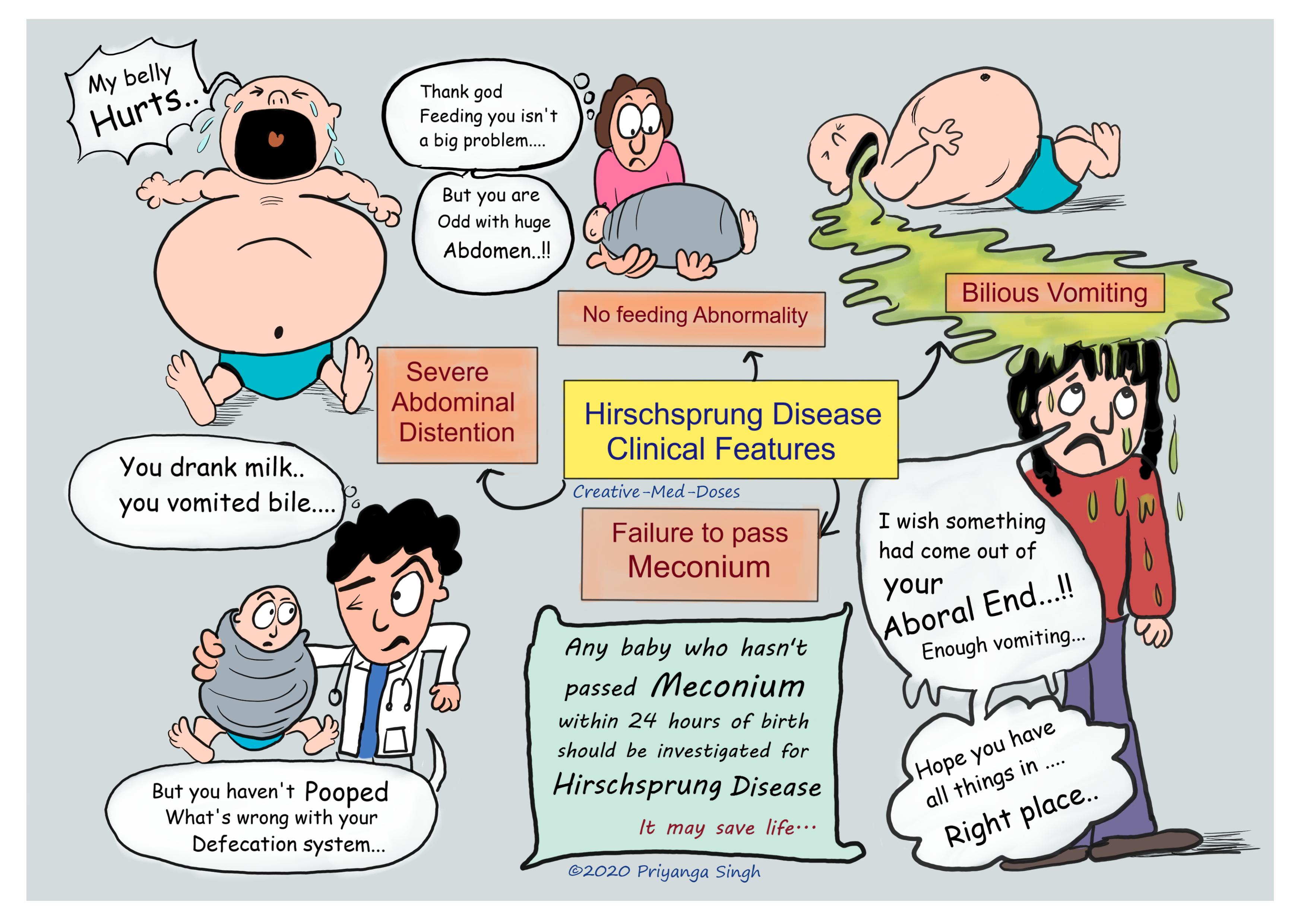
Hirschsprung Disease: Congenital aganglionic megacolon
Hirschsprung disease (HSCR) also called Congenital aganglionic megacolon is a motor disorder of the intestine, which is caused by the failure of neural crest cells to migrate completely during intestinal development during fetal life. The neural crest cells are (precursors of enteric ganglion cells). Their failure to migrate results in aganglionic segment of the colon, this lack of nerves causes constant contraction and causes functional obstruction.
Pathogenesis
It most commonly affects the rectosigmoid region and is the most common cause of neonatal intestinal obstruction.
Defects in the differentiation of neuroblasts into ganglion cells and ganglion cell destruction within the intestine can also contribute to the disorder, for example in Chagas disease amastigote destroy ganglion cells and leads to acquired Hirschsprung disease.
Genetics
RET is a gene that codes for proteins that assist cells of the neural crest in their migration through the digestive tract during embryogenesis.
EDNRB (endothelin receptor type B) gene codes for proteins that connect these nerve cells to the digestive tract. Thus, mutations in these two genes could directly lead to the absence of certain nerve fibers in the colon.
Macroscopically, the bowel in patients with HSCR can be seen to have a narrow aganglionic segment, a proximal transitional zone progressing from a narrow to a dilated lumen, and a dilated proximal portion of bowel (megacolon) The bowel wall is thickened as a result of hypertrophy of the muscular wall.
HSCR varies considerably in aganglionic length and classically affects the rectum and sigmoid in >70% of cases.
The functional abnormality results from the uncoordinated function of the aganglionic bowel and its failure to relax to allow forward propulsion of the intestinal contents. The functional obstruction always includes internal anal sphincter dysfunction and failure to relax.
Clinical features
- Severe abdominal distention
- Failure to pass meconium in first 24 hours- A delay in passage of meconium is the most common observation in the neonate suspected of having HSCR (>80%). Normal babies pass meconium within 24 hours, and even up to 48 hours. Any baby who passes no or little meconium within 24 hours of birth should be investigated for Hirschsprung disease. If this perinatal early diagnosis is missed, chances of newborn having Hirschsprung-Associated Enterocolitis increases many folds.
- Bilious vomiting
- chronic constipation
- Tight anal sphincter – digital rectal examination reveals tight anal sphincter and empty rectum. Stool and air (FART :P) gushes out explosively as soon as finger are removed (very characteristic).
- No stool in rectal vault.
- If patient has fever, diarrhea and abdominal distention suspect for obstructive enterocolitis which is most common cause of mortality in these cases.
...

...
Complications
- Hirschsprung-Associated Enterocolitis (HAEC)- Inflammation and infection of the intestine which leads to fever, foul-smelling and/or bloody diarrhea and abdominal distention. It can lead to Toxic Megacolon. It is the greatest cause of morbidity and mortality in children with Hirschsprung disease. Failure to recognize Hirschsprung disease in the early perinatal period places children at greater risk of HAEC
- Toxic megacolon and Sepsis- If not treated early, sepsis, transmural intestinal necrosis, and perforation are possible.
- Rupture/Perforation – peritonitis
- Volvulus
Diagnosis
Diagnosis is based on symptoms and confirmed by Rectal suction biopsy. Any baby who passes no or little meconium even after 24 hours should be investigated for Hirschsprung disease.
Abdominal X Ray –
It is initial test when diagnosis of HSCR is suspected and it shows findings of intestinal obstruction (HSCR is functional intestinal obstruction).
Findings:
- Decreased or absent air in rectum
- Dilated colon segment immediately proximal to aganglionic region
- Distal intestinal obstruction
Rectal biopsy (gold standard) – Full thickness (Biopsy under General anesthesia) or containing mucosa and submucosa (suction biopsy done at bed side).
Distally narrowed segment biopsy is done to extract mucosa and submucosa tissue samples. Lack of ganglion cells in submucosa is hallmark for Hirschsprung Disease. There are plenty of aberrant acetylcholine esterase (AChE) positive fibers in the lamina propria in a patient with Hirschsprung's disease. This increased AChE expression is associated with the hypertrophied extrinsic nerve fibers of the aganglionic segment in Hirschsprung's.
Normal nerves do not stain for AChE, but increased AChE expression is associated with the hypertrophied extrinsic nerve fibers of the aganglionic segment in Hirschsprung Disease.
The AChE-positive nerves in the aganglionic zone appear to correspond with the severity of the clinical presentation and degree of obstruction.
The histochemical determination of AChE in the rectal suction biopsy material eliminated the problem of inadequacy of suction biopsy specimens and the presence or absence of ganglion cells in the submucosa.
A number of studies reported a 99–100% diagnostic accuracy of HSCR using the criteria of increased AChE staining of nerve fibers in the lamina propria and muscularis mucosa, provided that an adequate submucosal specimen was obtained.
Contrast enema
Contrast (Barium) enema is performed to localize and determine the length of the aganglionic segment prior to surgery.
Findings:
- Transition Zone is seen funnel like dilated segment and contracted distal segment is seen.
- Mucosal irregularities
- Delayed evacuation of contrast
Anorectal manometry
It is used to measure contractility in the anus and rectum. This technique uses a balloon in the rectum to distend the rectum and a pressure sensor at the internal anal sphincter to measure the presence or absence of the rectosphincteric reflex.
In this disease, there is an absence of the rectosphincteric reflex that should relax the internal anal sphincter upon distension of the rectum.
Treatment
Surgery- Definitive management for the disease is surgery, using the “pull-through” method.
Treatment of enterocolitis includes nasogastric decompression, intravenous fluids, and antibiotics (metronidazole ± ampicillin and gentamicin).
Revision for today Henoch Schonlein Purpura
Subscribe YouTube channel Creative-Med-Doses
case scenario
A mother brings the 2-day-old male infant with abdominal distension and bilious vomiting to the pediatrician. She also informs pediatrician that he has not yet had a bowel movement. A contrast enema shows a transition point at the transverse colon between dilated ascending colon and non-distended distal portion of the colon. Which of the following is the most likely to confirm the diagnosis?
- Ach esterase staining on colonic Biopsy
- Rectal suction biopsy
- Colonoscopy
- CT scan
Answer is 1. Ach esterase staining on colonic Biopsy
Rectal suction biopsy is gold standard diagnostic method for Hirschsprung disease.
AchE staining done on frozen section from biopsy specimen taken via Rectal suction biopsy is confirmatory.