Hepatocellular Carcinoma (HCC)
- Hepatocellular carcinoma (HCC) is the most common primary liver malignancy affecting hepatocytes.
...

…
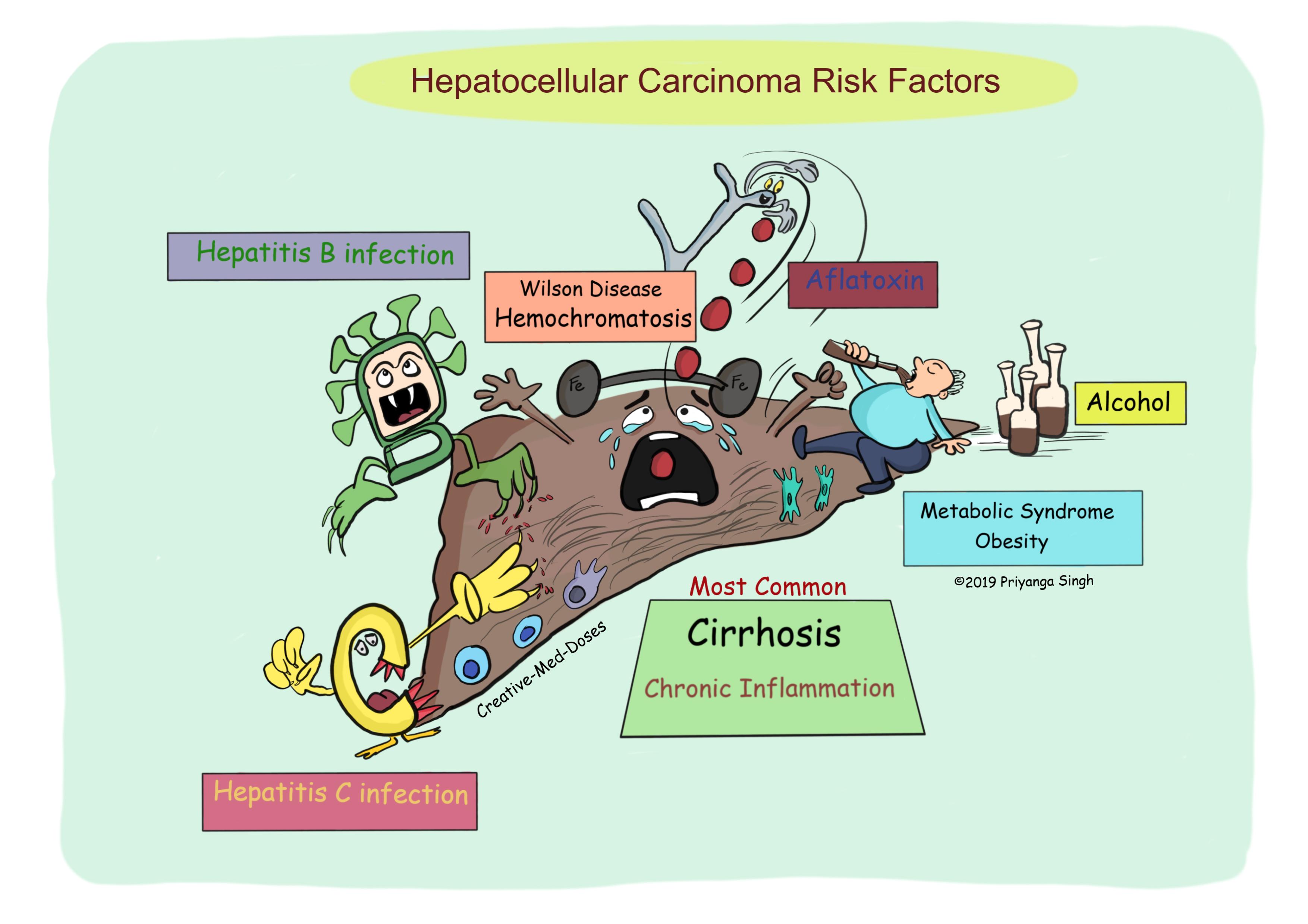
Risk factors
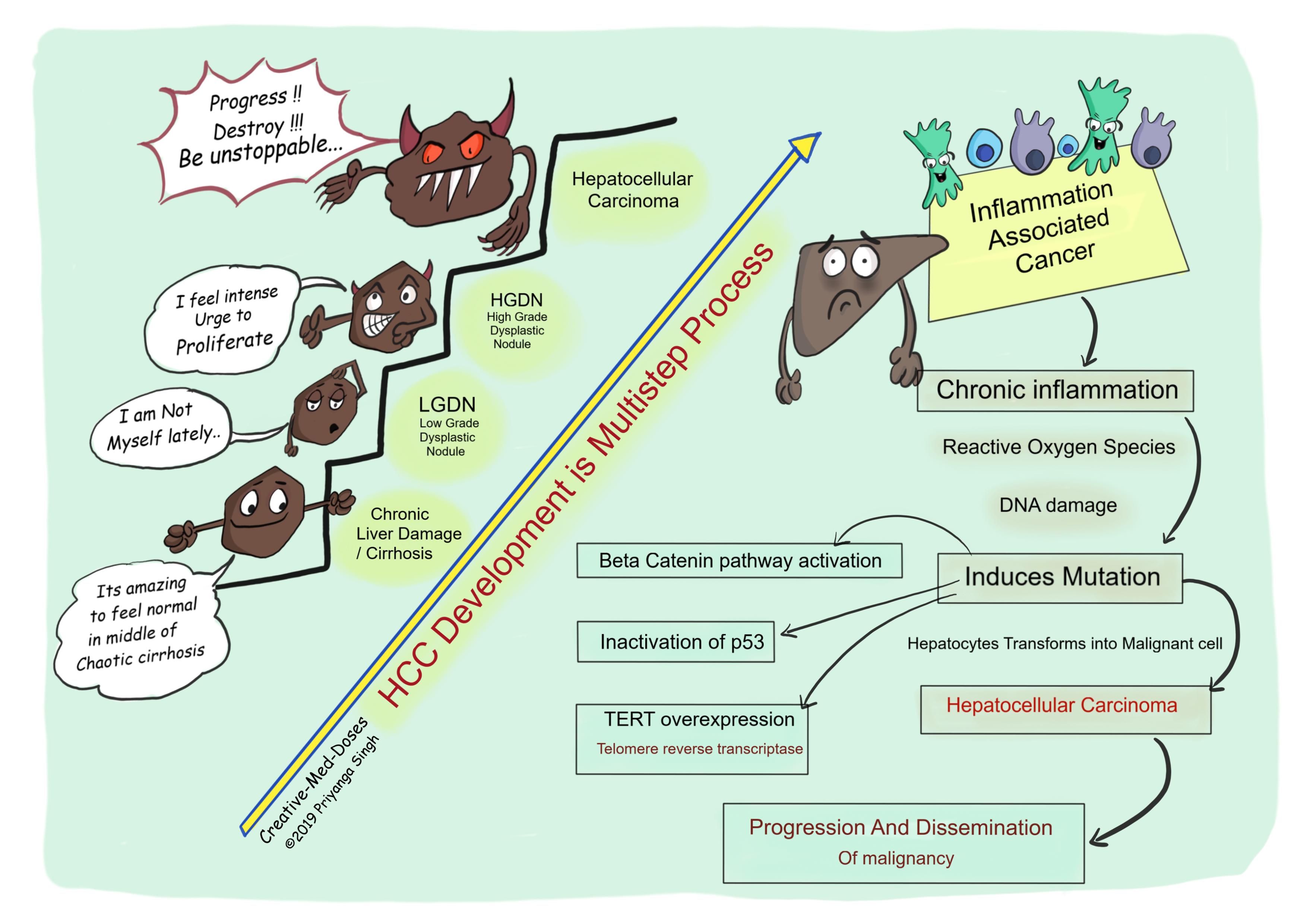
HCC is a prototypical inflammation-associated cancer, and inflammation causes immune microenvironment and oxidative stress to Induce DNA damage which plays pivotal roles in inducing mutations.
- Cirrhosis (most common cause)
- Chronic hepatitis B infection
- Chronic hepatitis C infection
- Alcohol abuse
- Metabolic Syndrome
- Hereditary Hemochromatosis, Wilson Disease
- Aflatoxin, produced by Aspergillus species
…

…
Molecular Pathogenesis
- HCC development is a multistep process it starts with precancerous cirrhotic nodules also called low-grade dysplastic nodules (LGDN) that evolve to high-grade dysplastic nodules (HGDN) that can transform into early-stage HCC.
- Molecular studies suggest that adult hepatocytes as the cell of origin, where molecular aberrations occur and accumulate to transform the hepatocyte into malignant cell.
Most Common Molecular Aberrations
- TERT (TElomerase Reverse Transcriptase) overexpression
- Inactivation of p53 and alterations of cell cycle
- Wnt/β-Catenin pathway activation
…

…
Clinical Presentation
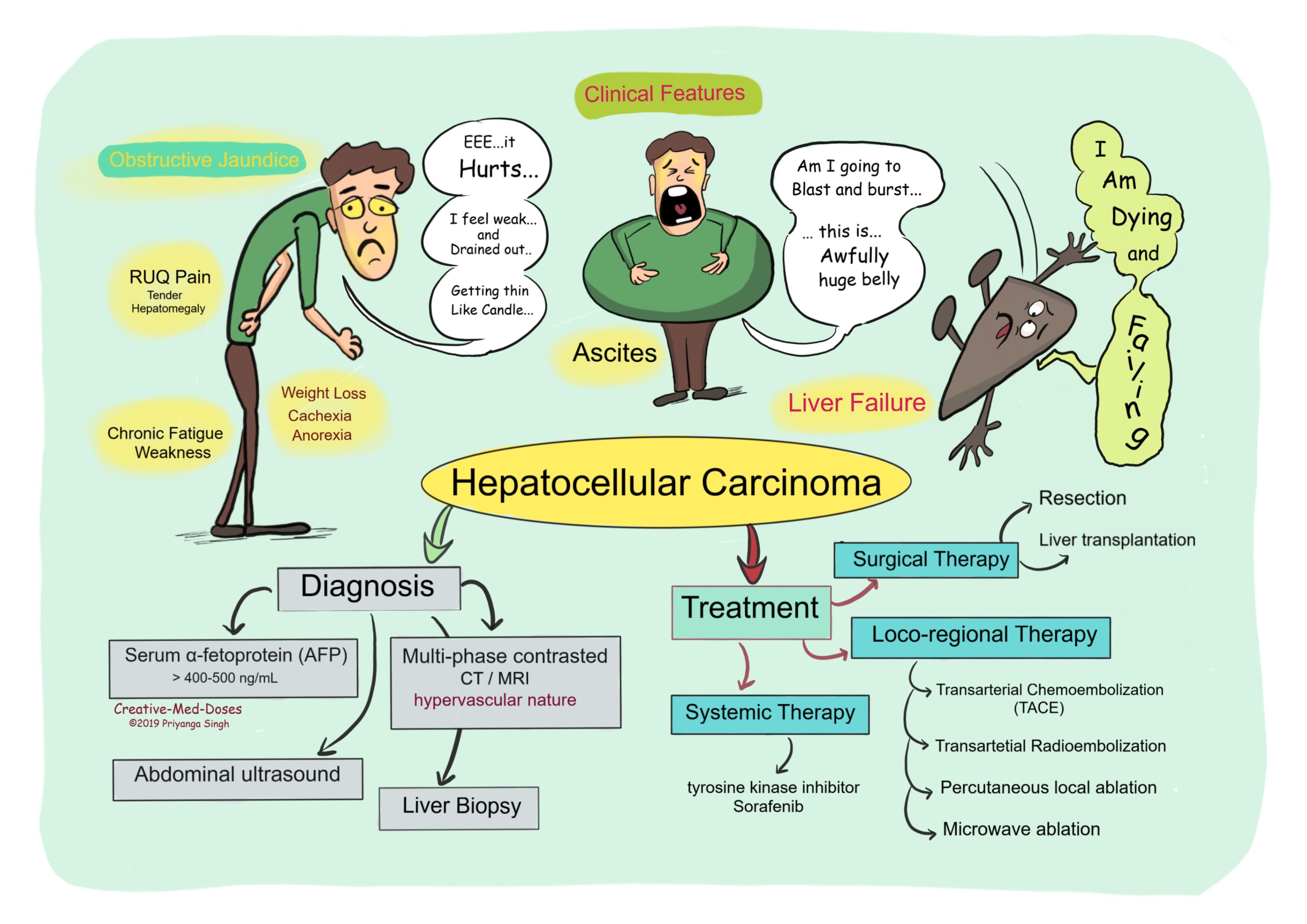
- Weight loss, cancer cachexia
- Anorexia
- Chronic Fatigue, weakness
- Tender Hepatomegaly (Right Upper Quadrant Pain)
- Obstructive Jaundice
- Ascites
- Liver Failure
- Sometimes patients can be asymptomatic and be incidentally found to have HCC due to routine screening in patients with cirrhosis
…

…
Diagnosis
- Serum α-fetoprotein (AFP) > 400-500 ng/mL is highly suggestive of HCC.
- Abdominal ultrasound- It is used as a screening test in patients with cirrhosis for development of HCC, irregular mass with poorly defined margin are suggestive of HCC.
- Multi-phase contrasted CT abdomen and Multi-phase contrasted MRI abdomen are confirmatory tests – In contrast-enhanced imaging techniques, the typical radiological hallmark of HCC consists of vascular uptake of the nodule in the arterial phase with washout in the portal venous or delayed phases. This radiological pattern captures the hypervascular nature of nodule which is characteristic of HCC.
- Liver Biopsy- is needed when radiology is not typical, and patient’s clinical presentation gives high suspicion for having HCC.
Treatment
- In most cases, the diagnosis of HCC is made when it is at advanced stage when patients have become symptomatic and have significant liver impairment. At this late stage, there is virtually no effective treatment that would improve survival. In addition, the morbidity associated with therapy is unacceptably high. Early diagnosis and treatment are the key for increased survival rates.
Following are treatment modalities -
Surgical Therapy
- Resection
- Liver transplantation
Loco-regional Therapy
- Trans arterial Chemoembolization (TACE) - high-dose chemotherapy is delivered (e.g., cisplatin and doxorubicin) to local areas in the liver, since it is localized it decreases the risk of developing systemic toxicities.
- Trans arterial Radioembolization- Transartetial radioembolization is a form of catheter-directed internal radiation that delivers small microspheres with radioisotopes directly into the tumor.
- Percutaneous local ablation-RFA (Radioactive Frequency Ablation) is the treatment of choice for local destruction of liver tumors. RFA produces coagulative necrosis of the tumor while leaving a safety margin around the tumor, leading this to be the most common local ablative therapy.
- Microwave ablation
Systemic Therapy-
- It is used for advanced disease
- Oral multitargeted tyrosine kinase inhibitor Sorafenib is the standard of care systemic therapy for HCC.