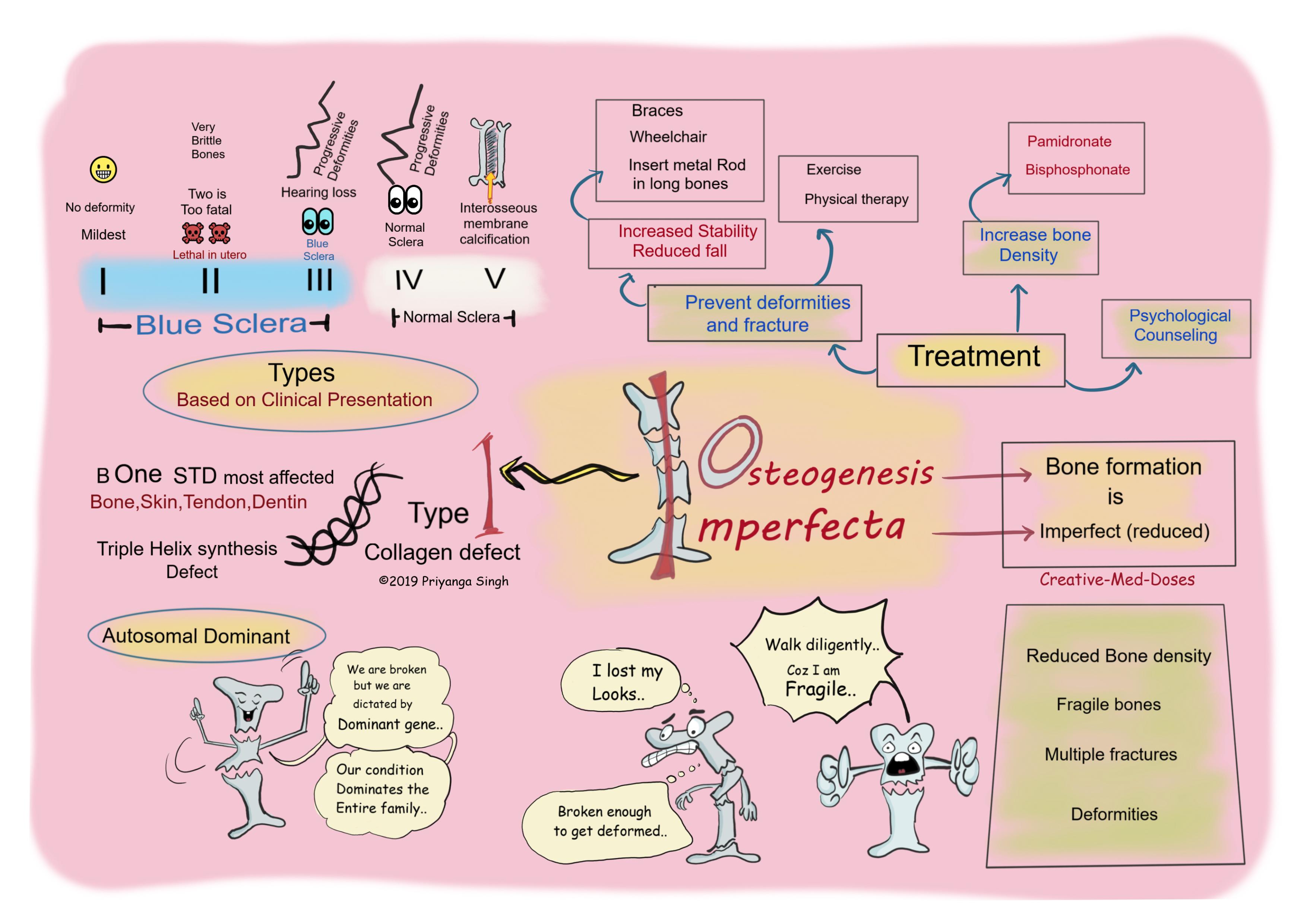
Osteogenesis Imperfecta: Brittle Bone Disease
- Osteogenesis Imperfecta is a hereditary connective tissue disorder with fragile bones due to decrease in the amount of normal Type I collagen.
- There is severe reduction in bone density that makes bone brittle, recurrent fractures with minimal trauma is very characteristic feature.
Pathogenesis
- Most common form is autosomal dominant. The mutation compromise triple helix folding, causes retention of mutant trimer in endoplasmic reticulum and degradation of misfolded protein. There is reduction in Type 1 collagen synthesis which leads to defect in bone formation, dentin formation and connective tissue defects in other parts of the body.
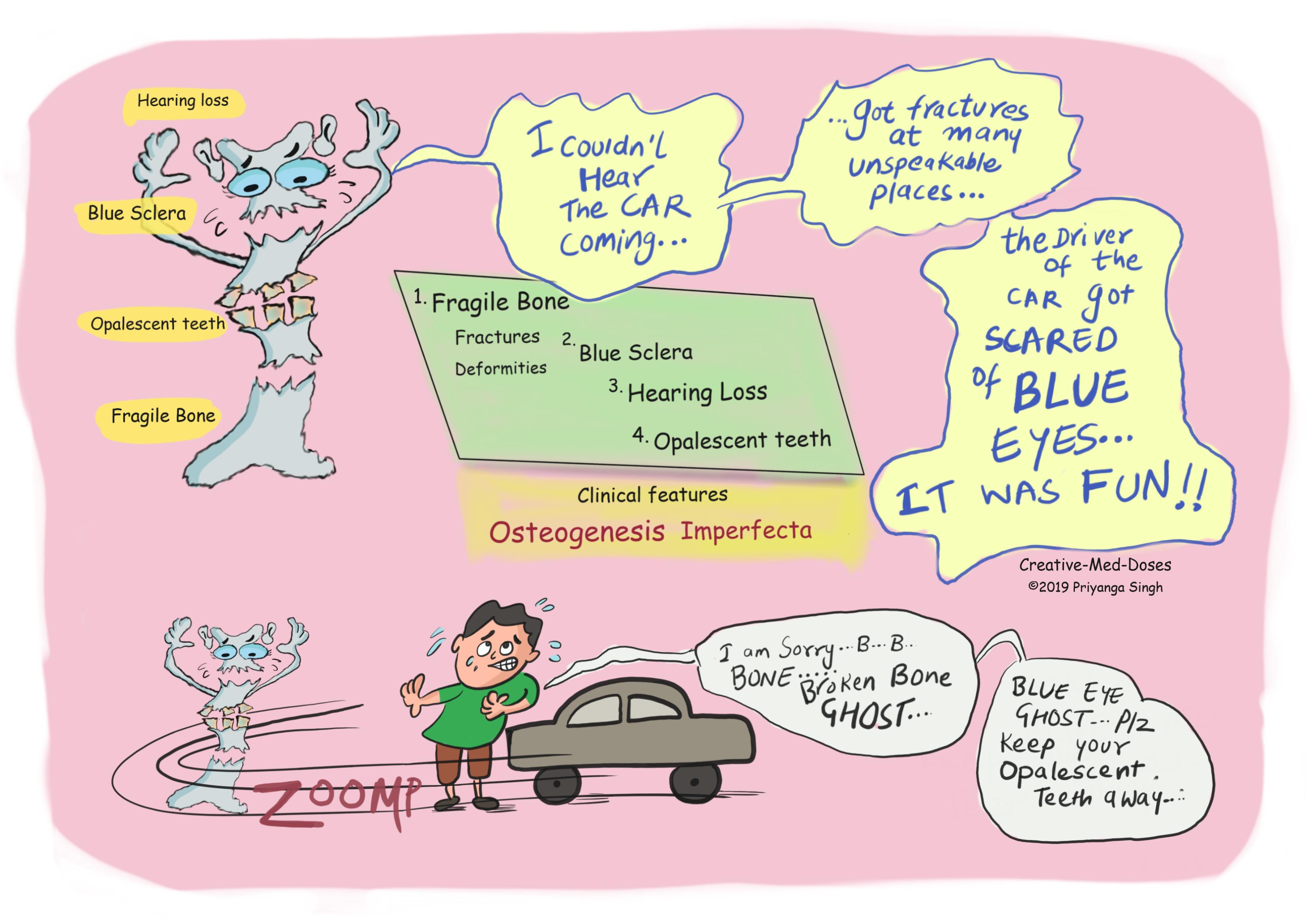
Clinical manifestation
- Skeletal defects
- Low Bone density
- Fragile bones
- Recurrent fractures
- Deformity
- Kyphoscoliosis
- Nerve root Compression
- Blue sclera – most forms have grayish blue sclera due to translucency of connective tissue over choroidal veins. (clue--veins are blue)
- Dentinogenesis Imperfecta- dental abnormalities, brown, opalescent teeth and teeth fractures due to defective dentin formation.
- Hearing loss- due to defective ossification of ear ossicles, it can be conductive, sensorial and mixed.
...
...
Classification
Based on clinical presentation
- Type I- mildest, no deformity
- Type II- lethal in utero, very fragile bones
- Type III- progressive deformity, hearing loss and blue sclera
- Type IV- progressive deformity, hearing loss and normal sclera
- Type V- interosseous membrane calcification and normal sclera
Diagnosis
- Clinical features and family history are enough in most cases, genetic analysis can be done to confirm the diagnosis.
Treatment
Target is to reduce fractures and deformity
- Physiotherapy and exercise
- Vitamin D and calcium supplements
- Insertion of metal rod into long bones to reduce fall
- Medicine to increase bone density
...
...
Case Scenario
- A 5-year-old boy slips on the floor and is rushed to the pediatrician. His work-up reveals a new fracture in his right femur, as well as multiple old fractures in his bilateral arms and legs at various stages of healing, his left leg is shorter than right leg. On physical exam, his sclera is blue. What is most likely diagnosis, suggest treatment options.
- Revision for today https://creativemeddoses.com/topics-list/collagen-synthesis-quick-review/
- Interesting video on Osteogenesis imperfecta https://www.youtube.com/watch?v=XBtZzny9qJQ