Hemochromatosis: Bronzed Iron Man
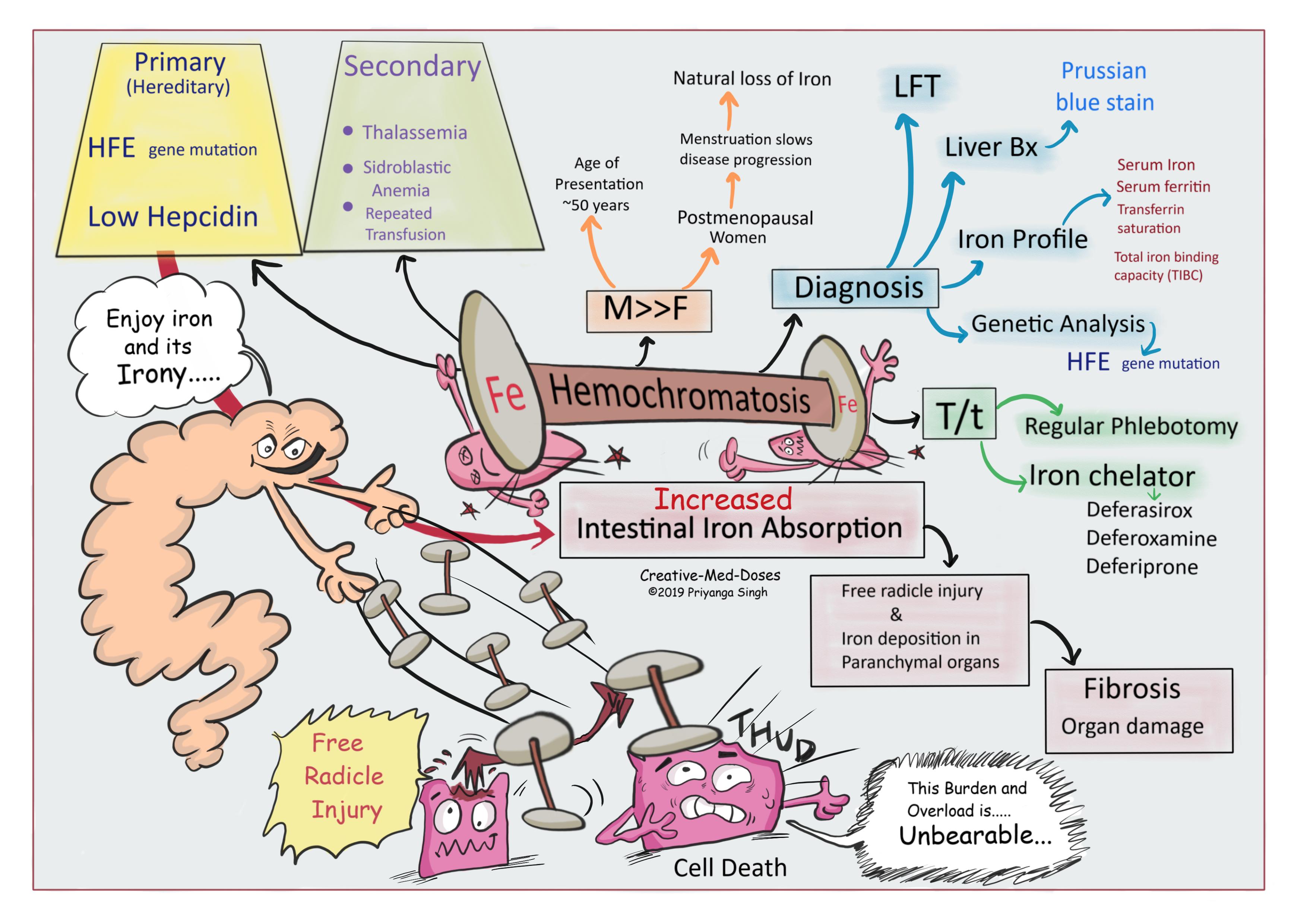
Hemochromatosis is an iron overload disease and excessive iron causes increased free radicle injury, fibrosis and organ failure. There is inappropriately low production of the hormone hepcidin. This leads to an increase in intestinal iron absorption and the deposition of excessive amounts of iron in parenchymal cells which leads to tissue damage and organ failure. Cirrhosis of the liver, diabetes mellitus, arthritis, cardiomyopathy, and hypogonadotropic hypogonadism are the major clinical manifestations of excessive iron load.
Men develop the disease earlier in their lifespan because they do not have the natural losses of iron like women during menstruation and pregnancy. Most women presenting with hemochromatosis are postmenopausal.
...

...
Types
Hereditary (Primary)- most often caused by a mutant gene HFE on chromosome 6. Persons who are homozygous for the mutation are at increased risk of iron overload.
Acquired (Secondary)- an iron-loading anemia, such as thalassemia or sideroblastic anemia, in which erythropoiesis is increased but ineffective causes excessive iron deposition in parenchymal tissues and end organ failure. Repeated blood transfusion in such cases aggravates the situation.
...

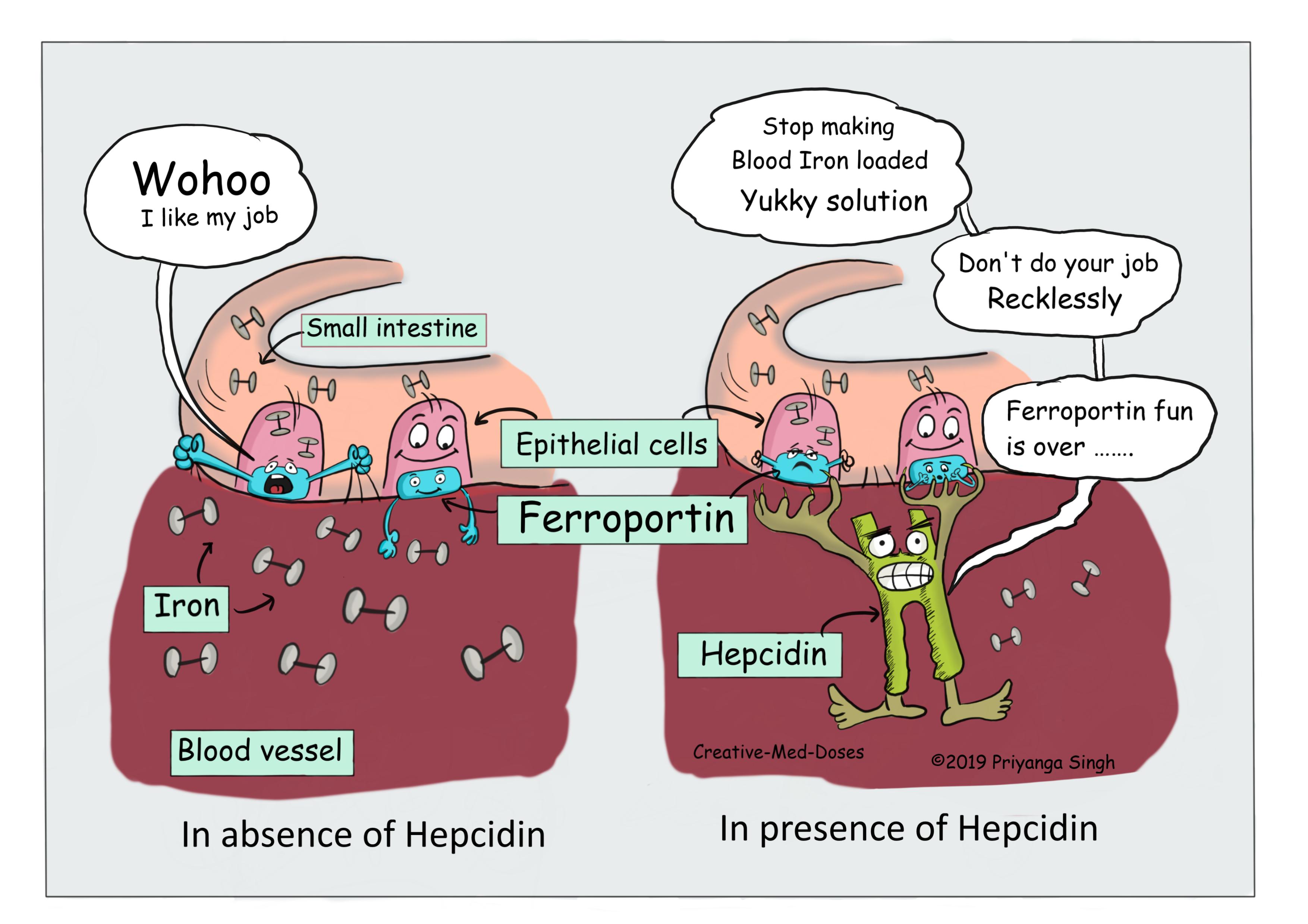
(Hepcidin reduces intestinal iron absorption by controlling ferroportin)
...
Clinical Manifestation
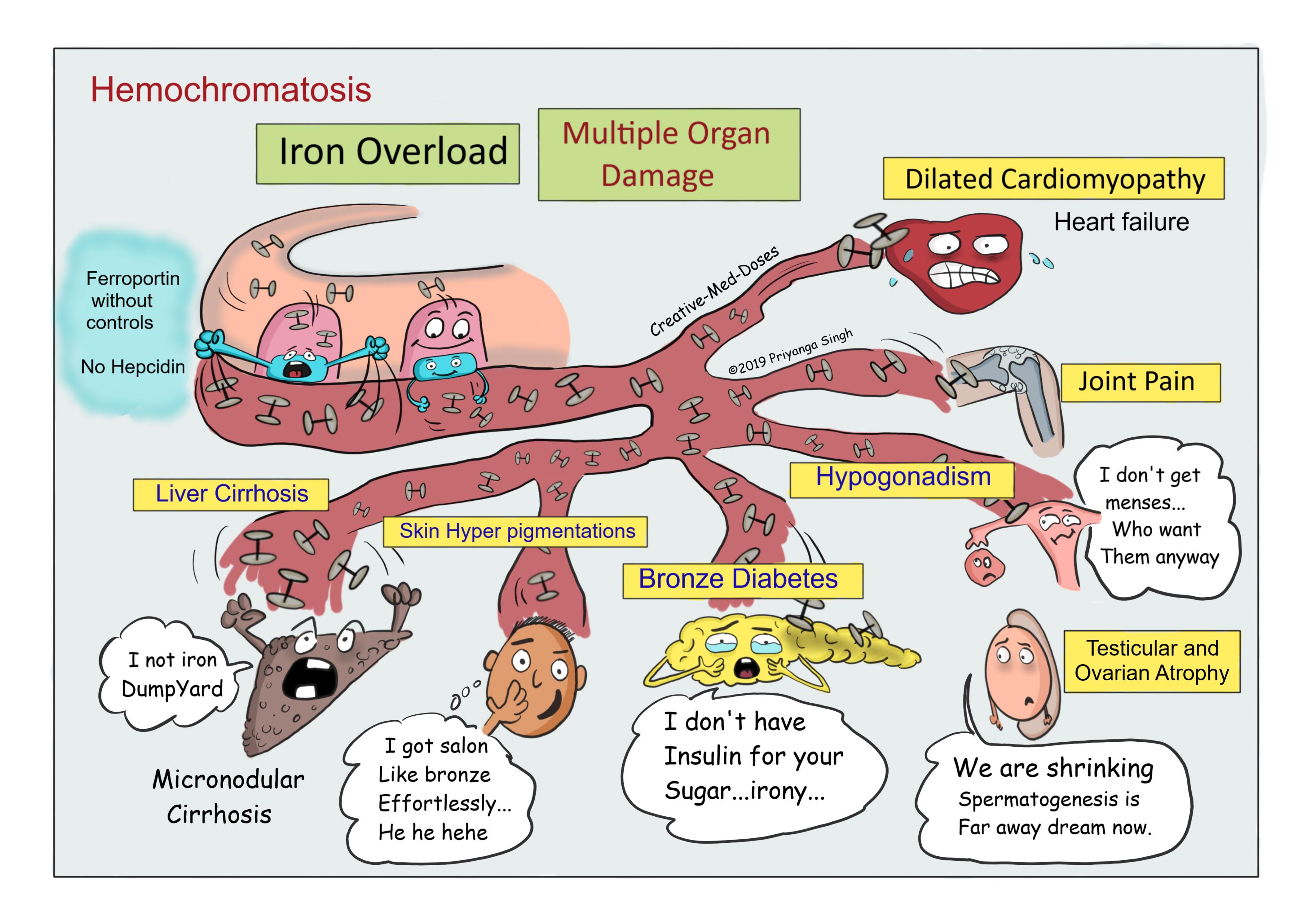
Liver (micronodular) cirrhosis – Patients present with abdominal pain, hepatomegaly and raised liver function tests. Hepatocellular carcinoma incidence is higher in cases with cirrhosis.
Skin hyperpigmentation- The characteristic metallic or slate-gray hue also called bronzing which results from increased melanin and iron in the dermis is frequently seen.
Diabetes mellitus- due to direct damage to pancreatic islet cells with iron deposition and reduced insulin production.
Arthropathy- excessive hemosiderin deposition in the synovial linings leads to synovitis. There is excessive deposition of calcium pyrophosphate, it damages articular cartilage which produces disabling polyarthritis.
Cardiac involvement- dilated cardiomyopathy, congestive heart failure and cardiac arrhythmia is frequent manifestation of iron deposition and subsequent organ failure.
Hypogonadism- it is present in both sexes and presents as loss of libido, impotence, amenorrhea, testicular atrophy, gynecomastia, and sparse body hair. It is result of decreased production of gonadotropins due to derangement of hypothalamic-pituitary function by excessive iron deposition.
Nonspecific – fatigue, weakness and malaise.
...

...
Diagnosis
The association of (1) hepatomegaly, (2) skin pigmentation, (3) diabetes mellitus, (4) heart disease, (5) arthritis, and (6) hypogonadism suggest the diagnosis.
Liver biopsy- shows signs of cirrhosis and deposited iron stains blue with Prussian blue stain.
Liver function test- liver enzymes are raised because of free radical injury and fibrosis.
Iron profile
- serum iron, serum ferritin and transferrin saturation are increased
- total iron binding capacity (TIBC) is low
Genetic testing- mutation in HFE gene is confirmatory
Treatment
Regular Phlebotomy
Iron chelator
- Deferasirox
- Deferoxamine
- Deferiprone
Case Scenario
A 60-year-old man presents to emergency department with exceptional dyspnea. He is known diabetic and takes insulin regularly. Physical exam reveals hepatomegaly and skin hyperpigmentation. His laboratory test reveals elevated serum iron, ferritin and reduced total iron binding capacity. What can be most likely cause –
- Chronic stress
- Liver cirrhosis
- Increased intestinal iron absorption
- Erythropoietin excess
Further reading http://www.scielo.org.co/pdf/rcg/v25n2/en_v25n2a12.pdf
Revision for today https://creativemeddoses.com/topics-list/polymyalgia-rheumatica-mournful-mornings/