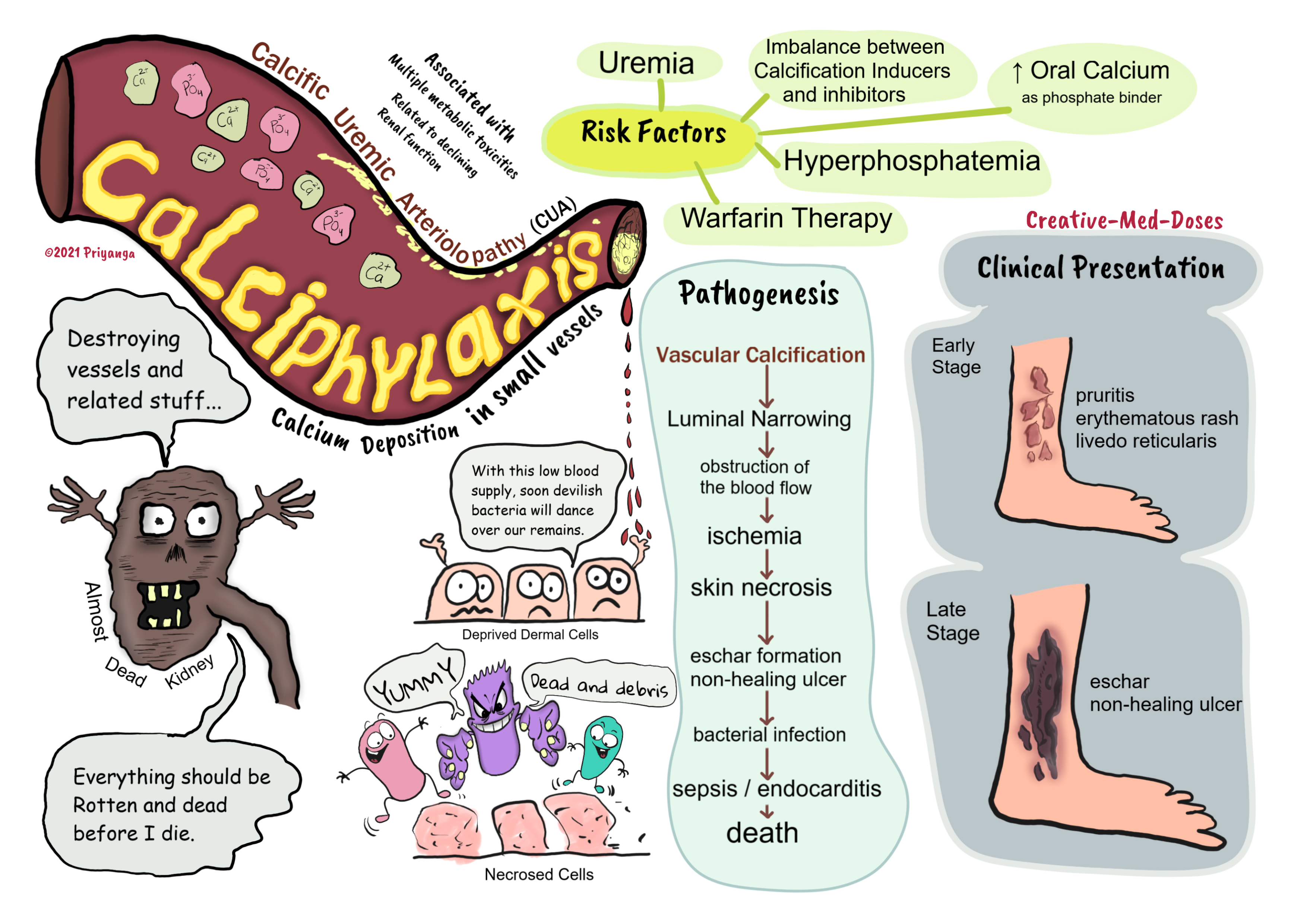
Calciphylaxis or calcific uremic arteriolopathy in CKD
Calciphylaxis or calcific uremic arteriolopathy (CUA) is a rare but fatal complication seen in patients in the late stage of chronic kidney disease.
The calcification of small vessels, especially arterioles, is a characteristic feature. And it eventually leads to the obstruction of the blood flow.
The blood flow obstruction causes ischemia, necrosis, severe pain, and color changes at the site of obstruction.
Pathophysiology
Calcific uremic arteriolopathy (CUA) has a multifactorial pathogenesis.
- The imbalance between inducers and inhibitors of calcification
- Increased use of oral calcium as a phosphate binder
- Secondary hyperparathyroidism
- Hyperphosphatemia – elevated serum phosphate induces a change in gene expression and switches vascular cells into osteoblast-like cells. These cells cause vascular calcification.
- Uremia in end-stage renal failure causes inflammation and suppression of calcification inhibitors. Suppression of calcification inhibitors leads to more calcification and obstruction.
- Warfarin in dialysis patients - Warfarin used in hemodialysis decreases the vitamin K–dependent regeneration of matrix GLA protein. The matrix GLA protein is crucial in preventing vascular calcification. That’s why warfarin treatment is considered a risk factor for calciphylaxis. If the patient develops calciphylaxis, discontinue warfarin and replace it with another anticoagulant.
The sequence of events in calciphylaxis vascular calcification→ luminal narrowing→ ischemia →skin necrosis and ulceration. If left untreated, it can cause secondary bacterial infection. Which can lead to sepsis, septic shock, and death.
Clinical presentation
In the early stage of Calciphylaxis or calcific uremic arteriolopathy, the patient presents with pruritus and cutaneous laminar erythema or a violaceous rash. It may resemble livedo reticularis.
In the late stage of disease, cases have painful eschars and painful non-healing ulceration and necrosis.
...

...
Diagnosis
CUA is a fatal complication of CKD, which demands timely diagnosis. It carries a high risk of mortality and various complications.
Following are helpful for early diagnosis -
- Clinical presentation and history of CKD
- Elevated serum phosphate and calcium levels
- Radiological image showing the calcification in tunica media of the arterioles and smaller subcutaneous vessels.
Skin biopsy
- A “skin biopsy” is a standardized diagnostic test.
- It is generally not recommended due to the high risk of poor healing or non-healing and infection.
Treatment
Conservative Therapy
- aggressive wound care (debridement, incision drainage)
- systemic antibiotics- to prevent secondary bacterial infection
- aggressively managing the renal disease with daily hemodialysis
- non-calcium containing phosphate binders
- Cinacalcet- calcimimetic agent which inhibits PTH release and accelerates wound healing
- Sodium thiosulfate- Sodium thiosulfate converts insoluble calcium in the tissues to soluble calcium thiosulfate. It improves the solubility of the calcium deposits and facilitating the removal of these deposits by mobilizing them. It promotes wound healing. It has antioxidant properties that protect against reactive oxygen species or free radical injury due to inflammation. It increases the formation of endothelial nitric oxide, which dilates the vessels. Vasodilation improves circulation and reduces the risk of ischemia.
Surgery
- It is not a favored line of treatment.
- Surgical stress can result in increased excitation of the sympathetic nervous system. It causes vasoconstriction and widespread necrosis.
- Surgery is for patients who are unresponsive to conservative treatment and have intractable pain.
Revision for today Selection of the Dominant Follicle and microenvironment - Creative Med Doses
Buy fun review books here (these are kindle eBook’s you can download kindle on any digital device and log in with Amazon accounts to read them). Have fun and please leave a review.