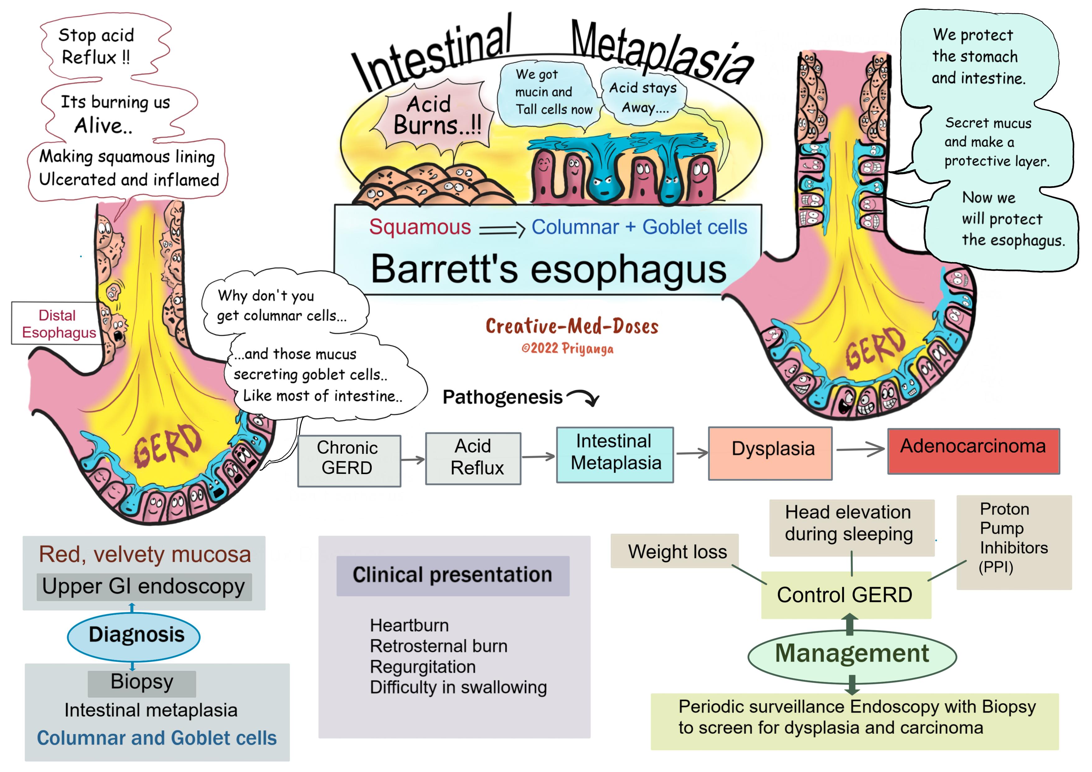
Barrett’s esophagus
Barrett’s esophagus is associated with chronic gastroesophageal reflux (GERD) and reflux esophagitis. Long-term acid exposure induces metaplastic changes in distal esophageal mucosa. And the esophageal squamous epithelium is replaced by columnar and goblet cells (intestinal metaplasia).
Pathogenesis
Chronic GERD → Increased acid exposure → Esophageal mucosal injury and squamous epithelial damage → Acute and chronic inflammatory changes → Reflux esophagitis → Recovery from damaged area involves metaplastic changes (Barrett’s esophagus) → Esophageal stem cells develop intestinal metaplasia (columnar and goblet cells) → Metaplasia may lead to dysplasia → Adenocarcinoma of the esophagus
Associated Risk Factors
- GERD
- Obesity
- Men > Women
Clinical Presentation of Barrett’s esophagus
- Heartburn
- Regurgitation
- Difficulty in swallowing
Diagnosis
History of chronic GERD and clinical presentation
Upper GI Endoscopy
Endoscopic examination shows tongues and patches of red, velvety mucosa extending upwards from the gastroesophageal junction. The normal esophageal mucosa looks smooth, pale, and pearly white.
Biopsy
Gastric and intestinal metaplasia is present in mucosa above the gastroesophageal junction. The presence of goblet cells with mucous vacuoles is defining feature of intestinal metaplasia. Goblet cells stain pale blue by H&E stain.
Management
GERD control
- Weight loss
- Head elevation during sleeping
- Proton pump inhibitors
Periodic surveillance endoscopy with biopsy to rule out dysplasia and adenocarcinoma
In case of Dysplasia / intramucosal carcinoma-
- Photodynamic therapy
- Radiofrequency ablation
- Endoscopic mucosectomy
- Surgical resection (esophagectomy)
....
....
Case scenario
A 55-year-old man presented to a clinician with chief complaints of retrosternal pain and difficulty swallowing. The upper GI endoscopy reveals red velvety mucosa alternating with pearly white esophageal mucosa. Which of the following cells are likely to be present in a biopsy from abnormal red and velvety mucosa?
- Keratinized stratified squamous epithelium
- Ciliated columnar epithelium
- Goblet cells with mucous vacuoles
- Cuboidal Epithelium
Revision for today Renal Hypoxia: Why kidney is prone to hypoxia and ischemic injury? - Creative Med Doses
Buy fun review books here (these are kindle eBooks you can download kindle on any digital device and log in with an Amazon account to read them). Have fun and please leave a review.
https://creativemeddoses.com/books/