Celiac Disease: Gluten Intolerance
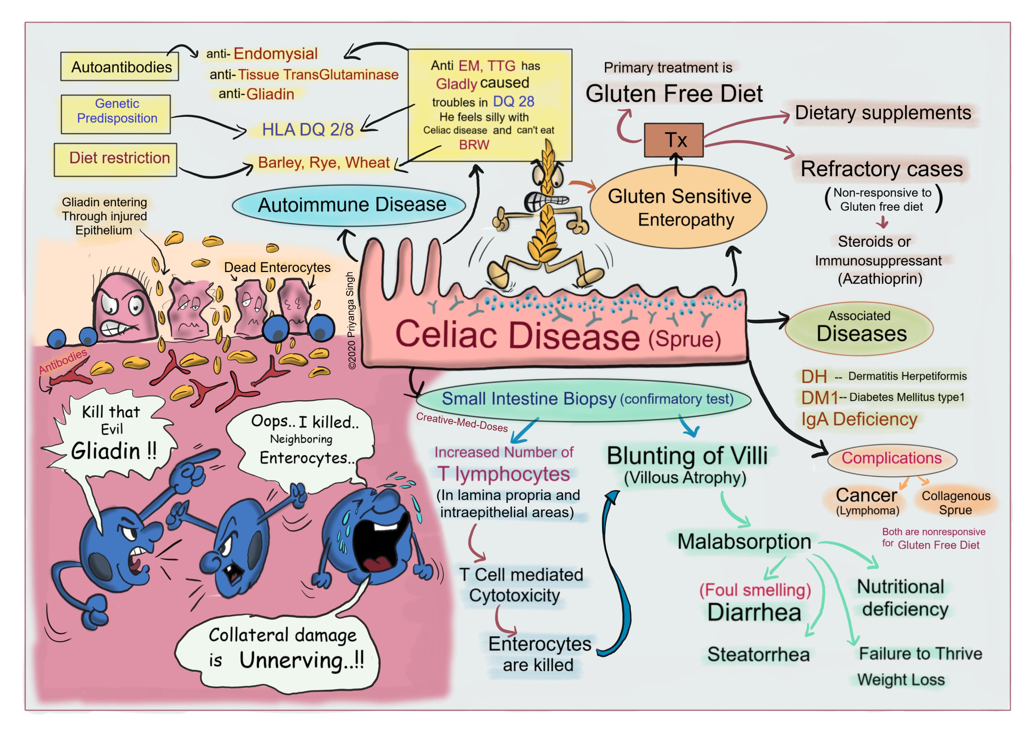
Celiac disease (Celiac Sprue) is an autoimmune disease caused by the ingestion of gluten, it is also called gluten sensitive enteropathy. Gluten is the protein component of wheat, rye and barley. There is an intestinal immune reaction to gluten in genetically predisposed individuals causing damage to intestinal epithelium.
Following three factors are working together in disease causation -
- Environmental factors- eating gluten containing food (BRW) -Barley, Rye and Wheat
- Immunologic factors- Presence of Autoantibodies, anti-Endomysial, anti-Tissue Transglutaminase (TTG) and anti-Gliadin.
- Genetic factors- associated with HLA-DQ2 or HLA-DQ8 alleles.
Pathogenesis
There are multiple proposed pathways involved with ingestion of gluten that leads to enterocyte destruction and subsequent villous atrophy.
Gliadin which is glycoprotein extract from the gluten is main culprit here—
- It is directly toxic to the enterocytes of genetically predisposed individuals.
- It causes overexpression of IL-15 by enterocytes which in turn triggers activation and proliferation of CD8 intraepithelial lymphocytes. These lymphocytes are cytotoxic and Kills enterocytes. Intraepithelial lymphocytes are increased in number.
- Gliadin upregulates stress molecule MIC-A on enterocytes and NKG2D receptors on intraepithelial lymphocytes of genetically predisposed individuals (HLA DQ2 / DQ8 alleles).MIC-A molecules are stress molecules, they interact with NKG2D receptors on natural killer cells and CD8 lymphocytes. It leads to cytotoxic killing of enterocytes and loss of villi.
- This loss of villi/villous atrophy leads to malabsorption of nutrients, diarrhea, steatorrhea and nutritional deficiencies.
- These activated lymphocytes stimulate B cells/ plasma cells to secret autoantibodies named anti-endomysial, anti-TTG and anti-gliadin.
...
...
Clinical presentation
Pediatric cases
Patient presents with classical symptoms of irritability, abdominal distention, diarrhea, failure to thrive and weight loss. Most cases present typically between 6 and 24 months of age with history of introduction of gluten in diet. Few cases present with Dermatitis herpetiformis, which has characteristic pruritic, papulovesicular lesions on shoulders, arms and cheeks.
Adult cases
Most cases present between 30 and 60 years of age. Anemia (iron deficiency), diarrhea, bloating, fatigue and weight loss are characteristic features.
Diagnosis
Diagnosis is based on clinical symptoms and confirmed with small bowel biopsy and positive serology.
Serology
It is best initial test. The most sensitive tests are the presence of IgA antibodies to tissue transglutaminase or IgA or IgG antibodies to deamidated gliadin. Anti-endomysial antibodies are highly specific but less sensitive than other antibodies.
Small intestine Biopsy
Biopsy specimens should be taken from the duodenum or proximal jejunum (small intestine), which are exposed to the highest concentrations of gluten.
The histopathology reveals-
- increased numbers of T lymphocytes, with intraepithelial lymphocytosis
- crypt hyperplasia
- villous atrophy (blunting of villi)
Intraepithelial lymphocytosis and villous atrophy can be present in other disorders, for example lactose intolerance, viral enteritis and inflammatory bowel disease. The combination of histologic and serologic findings is most specific for diagnosis of celiac disease.
Associated Diseases
- DH (Dermatitis Herpetiformis)- itchy papulovesicular lesions responsive to dapsone.
- DM1- Diabetes Mellitus type1
- IgA deficiency
Complications
- Development of cancer
- Collagenous Sprue
Treatment
- Gluten free diet is central measure.
- Refractory cases are treated with steroids or immunosuppressive agents (Azathioprin).
Further reading https://www.ncbi.nlm.nih.gov/pmc/articles/PMC2775561/
Revision today https://creativemeddoses.com/topics-list/hereditary-spherocytosis-intrinsic-hemolysis/
Buy fun review books here (these are kindle eBook’s you can download kindle on any digital device and login with Amazon accounts to read them). Have fun and please leave review. https://creativemeddoses.com/books/
Case scenario
A 2-year-old girl is brought by mother to the pediatrician for diarrhea and intermittent vomiting. She has been passing 5-7 loose stool per day for last 7 months. On physical examination several red blistering excoriated areas are noticed over cheeks and shoulders. Her parents have eliminated dairy from her diet, but symptoms are persistent. Which of the following is most likely finding on intestinal biopsy?
- A. Crypt abscess and crypt distortion
- B. Non caseating granuloma in colon
- C. Atrophy and blunting of villi
- D. Lymphocytic infiltration and epithelial cell apoptosis